Cone Beam Imaging Is Here and Now!
 Paul Feuerstein my Dental Technology Solutions partner sent me this image from Conebeam.com
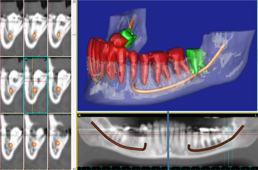
Conebeam imaging is here and getting more routine then you might think.
Case History: Patient presented with a complaint of pain around tooth #17. Clinical diagnosis was pericoronitis. When panoramic x-ray revealed a close association between the apices of #17 and the mandibular canal, the oral surgeon ordered a CT scan. From the CT it appeared that the mandibular canal passed between the roots of #17. The 3D reconstruction clearly shows this to be the case.
Go check out the Conebeam.com web site and sign up for their newsletter.

Comments